Electron Microscopy
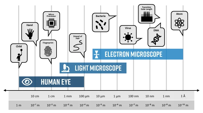
Electron microscopy (EM) is a technique for obtaining high resolution images of biological and non-biological specimens. It is used in biomedical research to investigate the detailed structure of tissues, cells, organelles and macromolecular complexes. The high resolution of EM images results from the use of electrons (which have very short wavelengths) as the source of illuminating radiation. Electron microscopy is used in conjunction with a variety of ancillary techniques (e.g. thin sectioning, immuno-labeling, negative staining) to answer specific questions. EM images provide key information on the structural basis of cell function and of cell disease.
Reserve SEM/TEM Instruments and Training
For access to our Scanning Electron Microscopes (SEM) and Transmission Electron Microscopes (TEM), please visit our Core Facility Website .Here, you can:
- Make Reservations for SEM and TEM instruments.
- Register for Training sessions to enhance your skills in using these advanced microscopy tools.