Analysis of Small Microplastics and Nanoplastics in Water and Food Matrices / AMiNaWa
Objectives
- Optimization, further development and application of Raman microspectroscopy (RM) for the detection, identification and quantification of small microplastic (MP) particles
- Improvement, validation and testing of an automated Raman-based method for the analysis of MP particles between 1-5 μm and smaller than 1 μm
- Development and optimization of methods for the separation of microplastics from food matrixes and their subsequent analysis
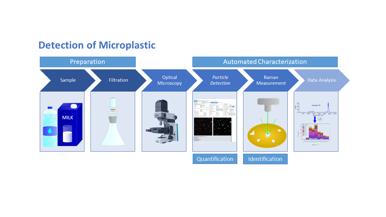
Methods of Approach
- Raman Microspectroscopy (RM)
- Image analysis employing TUM-ParticleTyper 2
- Field-flow fractionation (FFF)
- Scanning electron microscopy (SEM)
Description
Public concern is growing over the ingestion of microplastics and nanoplastics through consumables like drinking water and milk, and its potential impact on human health. To initiate a comprehensive food safety risk assessment for these particles, reliable information on the prevalence of commonly encountered small microplastic and nanoplastic types (e.g., PA, PC, PE, PES, PLA, PMMA, PP, PS, PTFE, PU, PVC, PBS, PHA) in various matrices is imperative. While Raman microspectroscopy is widely regarded as the gold standard for microplastic analysis, there is a scarcity of studies focusing on the analysis of small microplastics within the 0.5 to 20 μm range. Consequently, a dependable method for their quantitative analysis in terms of size, quantitiy and chemical composition is currently missing.
The overarching goal of this project is to evaluate and enhance existing techniques, such as Raman microspectroscopy, scanning electron microscopy (SEM), and field-flow fractionation (FFF), to detect and identify micro and nanoplastics in water and milk matrices down to 100 nm. For particles down to 5 μm the current TUM-ParticleTyper 2, a Python-based software tool that determines particle location and shape from microscopic images of a filter surface and subsequent analysis of Raman measurements, will be refined. The Raman measurement parameters will be optimized for the automation program, assessing the feasibility of measuring down to 0.5 μm using this method. Its usability for particles down to 1 μm has already been established. An alternative approach for measuring particles ranging from 5 μm down to 100 nm involves an online-coupled combination of Raman microspectroscopy and field-flow fractionation (FFF), in series with a multiangle light scattering (MALS) detector and a UV detector. Optical trapping with the same laser ensures that particles remain in the laser focus for spectral acquisition.
Challenges in this endeavor include the escalating number of particles in smaller size ranges, resulting in increased analysis time, as well as the agglomeration of particles and low concentrations. Efforts will be directed toward improving the deagglomeration of detected particles through software enhancements. Another challenge involves minimizing foreign matrix interference to reduce the appearance of non-plastic particles. Therefore, optimization of sample preparation methods is crucial to diminish foreign matter.