Raman Microspectroscopy (RM)
Basics
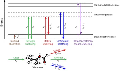
Raman Spectroscopy is based on the effect of inelastic light scattering due to changes in the polarizability of electrons and associated with measuring of molecular vibrations. This method provides “fingerprint” spectra that are unique to each specific compound and contain information on vibrations of molecules or groups of atoms and hence on chemical composition and structure of samples. Moreover, the coupling of Raman Spectroscopy with microscopy (Raman Microspectroscopy) enables for chemical analysis with high spatial resolution (below ~ 1 µm) and sensitivity. Furthermore, since water is a weak Raman scatter, in situ and iv vivo analysis of (micro)biological samples can be performed.
Advantages of Raman Microspectroscopy
- RM provides fingerprint spectra
- No or little sample preparation
- Non-contact and non-destructive
- No staining is necessary
- No or little interference from water
- Spatial resolution in µm-range
- Raman mapping/chemical imaging
Surface-Enhanced Raman Scattering (SERS)
Due to the low quantum efficiency of the Raman effect (typically 10-6–10-8), RM suffers from limited sensitivity. However, Raman scattering can be significantly enhanced if a molecule is attached, or in immediate proximity, to nanometer-roughened metal (Ag, Au, or Cu) surfaces. This effect, known as surface-enhanced Raman scattering (SERS), leads to Raman signal enhancements in the range of 103–106. Under certain conditions (at “hot spots” – closely spaced particles or rough nanostructures), enhancement factors up to 1011 (sufficient for single-molecule sensitivity) can be achieved. At least two effects contribute to the observed total enhancement. The electromagnetic enhancement effect is based on “localized surface plasmon resonance”, that takes place on the nanometer scale of SERS substrates, while chemical enhancement or “charge transfer”, is assumed to involve an electronic coupling between the adsorbed analyte and metallic substrate.