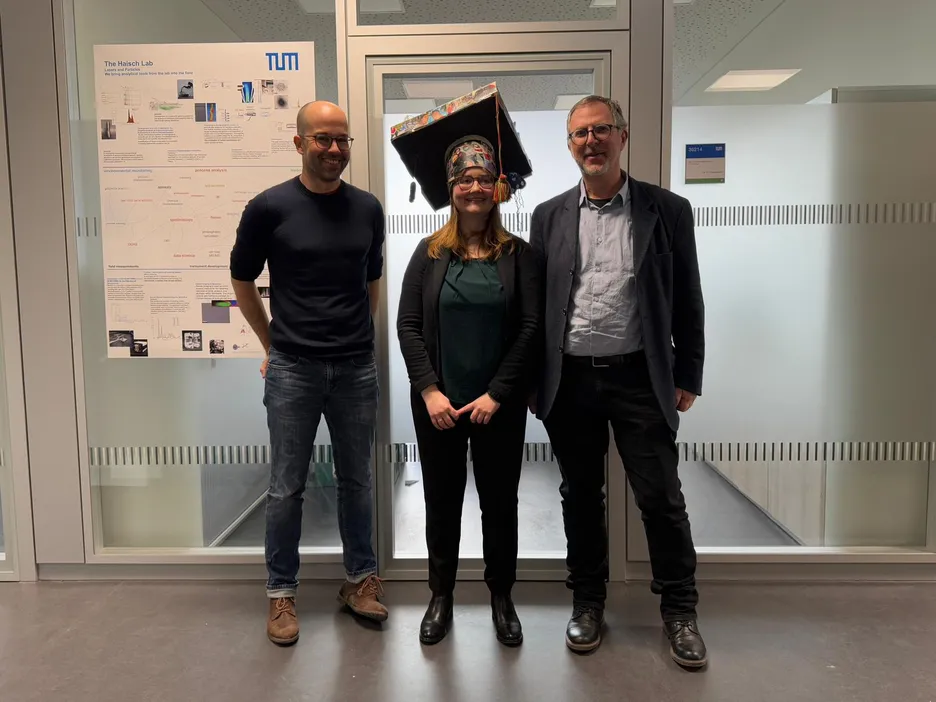
Julia Neumair successfully defended her PhD-Thesis-Congratulations!

Affinity-based methods for the isolation and detection of extracellular vesicles, bacteria and biomarkers in body fluids
Abstract
Affinity describes a specific, non-covalent, and therefore reversible interaction between two partners; an affinity binder and its target. One well-known example are antibodies and their antigens. Affinity-based analysis and separation techniques are applied to isolate and detect various analytes like small molecules, proteins, or even cells or bacteria. The presence of specific analytes like biomarkers or bacterial cells in various body fluids can give hints for infections, sepsis, or other diseases. In this thesis, three different approaches were performed to show the application of affinity-based detection and isolation methods for three different analytes from three different body fluids.
For the isolation of extracellular vesicles (EVs) from urine a novel monolithic immunofiltration assay using nanobodies was developed. EVs are small membrane-enclosed vesicles released from cells. Being carriers of cellular information such as proteins, DNA or RNA, EVs and their cargo can be used as biomarkers for different diseases like cancer or neurodegenerative diseases. Traditional isolation methods focusing on size-dependent discrimination like filtration, ultracentrifugation or asymmetrical flow field-flow fractionation, unfortunately lack the ability to discriminate between the cell types the EV origin from. Immunoaffinity-based methods can overcome this problem. Single domain antibodies with high antigen affinity, so called nanobodies, were used as affinity binders. Nanobodies targeting the surface-associated protein CD63 were immobilized indirectly via another nanobody-antigen interaction on a macroporous epoxy-based monolith with pore sizes of 22.4 ± 8.8 µm. After EV capturing, elution was carried out first competitive followed by pH-dependent elution. With this nanobody-based monolithic immunofiltration it was possible for the first time to isolate roughly 3 × 10¹⁰ EVs in two different size categories of about 36 and 130 nm from 7.5 mL of urine.
Infections in body fluids are critical conditions, that have to be treated rapidly and appropriately. In some cases, this is not easily done, like for infected ascites. Ascites is a life-threatening complication of diseases like liver cirrhosis, leading to a large fluid accumulation in the abdominal cavity. Infection with pathogenic bacteria like Escherichia coli or Enterococcus faecalis is not only one of the biggest causes of ascites-related death but also difficult to diagnose by culture due to very low bacterial concentration. Molecular methods are faster, but more expensive and labor intensive. Here affinity-based enrichment can help to facilitate identification and treatment. Crucial for effective affinity filtration that can facilitate diagnosis through culture are a combination of selective affinity binders and desorbing acting elution buffer. But their interactions are unfortunately not always easily predictable, which leads to a high number of tests that have to be performed to determine both components. The use of bioanalytical screening platforms can herby reduce the work effort. Therefore, in this thesis, a flow-based chemiluminescence (CL) microarray screening assay was established on the automated analysis platform, the Microarray Chip Reader – Research (MCR-R). For binding verification of bacteria to immobilized affinity binders, living E. coli and E. faecalis cells were tagged with biotin. These labeled bacteria were able to bind to streptavidin horseradish conjugates for CL detection on MCR-R. Recoveries of living cells were found to be 98 ± 8.8% and 75 ± 28% for E. coli and E. faecalis, respectively. Multiple affinity binder candidates like antibodies or antibiotic acting molecules were immobilized in rows of 5 spots each onto a surface of polycarbonate foil-based microarray chips. Biotin-tagged bacteria were pumped through the microarray chip to interact with the bound affinity binders and detected with the CL assay. Manual flushing of the microarray chip with elution buffer and a second CL detection step allowed not only for simultaneous screening for affinity binders, but also for simultaneous screening of suitable elution buffers. While the best affinity binders were found to be antibodies against the respective bacteria, the antibiotic Polymyxin B was found as an affinity binder for both bacteria. The sugar methyl alpha-D-mannopyranoside was discovered surprisingly as a suitable elution buffer for Polymyxin B, found due to the simultaneous elution studies.
Although upper airways diseases caused by allergy or bacterial or viral infections must be treated differently, it can be hard to distinguish due to similar symptoms. Biomarkers present in nasal secretion like cytokines can be used for discrimination. To enable rapid and precise treatment, fast and simultaneous detection of various biomarkers is crucial. Flow-based microarray assays outdo traditional static detection methods in means of speed and simultaneous detection of multiple analytes. Therefore, a flow-based CL-SMIA (sandwich microarray immunoassay) for the quantification of interferon-beta was established on the MCR-R. Commercially available antibodies designed for a sandwich enzyme-linked immunosorbent assay (ELISA) were used for the assay development. Comparison to the ELISA showed comparable detection limits for ELISA and CL-SMIA with 1.60 pg mL-1 and 4.53 pg mL-1, respectively. While the ELISA took 5 h 40 min to complete, the results from CL-SMIA were obtained after 1 h 15 min. The comparison study showed that CL-SMIA is more efficient in time and cost for simultaneous detection of multiple analytes and fewer sample sizes than ELISA. While in this proof-of-principle study only one biomarker was used, it paves the way for future simultaneous analysis of multiple biomarkers in nasal secretion
Summarizing, in this thesis it was possible to show the great potential of affinity-based bioassays for detection and isolation of complex analytes from body fluids in three unique ways. The work presented can be used as a foundation for future developments of diagnosis in special body fluids.