Raman Microspectroscopy for Non-invasive, Three-dimensional Analysis; Part of LegioTyper Project
Objectives
- Spatially resolved analysis of biofilms by Raman microspectroscopy (RM)
- Spectroscopic detection of Legionella pneumophila inside of host cells
- Differentiation of intra- and extracellular Legionella cells
Method of Approach
- Confocal Raman microspectroscopy (CRM)
- Surface-enhanced Raman spectroscopy (SERS)
- Stable isotope Raman microspectroscopy (SIRM)
- Cryo-scanning electron microscopy (CSEM)
Description
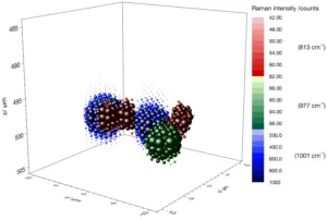
Raman spectroscopy (RS) has gained vast application possibilities since the discovery of lasers and high-performance detectors. Generally, solid as well as liquid and gaseous samples are measurable by RS. As a vibrational spectroscopy technique, it provides comprehensive information about the molecular structure of substances and is a non-destructive analytical method. Additionally it shows only minor interferences with water. A resolution down to the diffraction limit is possible with an integrated microscope. Raman microspectroscopy (RM) enables the chemical analysis of microscopic structures like single bacteria. Three-dimensional measurements may be executed if the sample is transparent in the region of the excitation wavelength.
General goal is to explore this 3D analysing capability of the confocal Raman microscopy (CRM). One application field is the analysis of microbial samples and especially biofilms. These represent a dominating form of appearance for microorganisms. The basic structure of biofilms is formed by extracellular polymeric substances (EPS). The natural polymers are usually highly hydrated, rendering the biofilms sensitive to changes of environmental conditions. As a non-contact technique with minimal need of specimen preparation, RM is well suited to chemically visualize 3D structures of biofilms. In combination with the use of stable isotope approach comprehensive studies of this complex ecosystem are feasible.
In particular, the analysis of Legionella pneumophila containing biofilms is performed. This bacterium is the causative germ of the Legionnaires’ disease, a pneumonia with a high lethality. Aim of this part of research (LegioTyper project) is to detect Legionella inside of amoebas and differentiate them from extracellular bacteria. Legionella spp. are known to multiply in unicellular hosts and to outlast disinfection or even increase virulence if occurring intracellular. CRM is supposed to be applied as an analytical reference technique for biochemical methods like immunoassays to guarantee the detection of intracellular Legionella. The results may also enhance the understanding of the interactions between amoeboid hosts and L. pneumophila as well as its pathogenic properties.
Publication
R. Weiss, M. Palantinszky, M. Wagner, R. Niessner, M. Elsner, M. Seidel & N. P. Ivleva, Surface-Enhanced Raman Spectroscopy of Microorganisms: Limitations and Applicability on the Single-Cell Level. Analyst 2019 doi.org/10.1039/C8AN02177E
Posters
R. Weiß, R. Nießner, M. Seidel, N. P. Ivleva, Auswirkung physiologischer Bedingungen auf Detektion von Mikroorganismen mittels Oberflächen-verstärkter Raman-Spektroskopie, WASSER 2018, 07.-09.05.2018, Papenburg, Deutschland.
R. Weiß, R. Nießner, M. Seidel, N. P. Ivleva, Raman-Mikrospektroskopie für zerstörungsfreie, dreidimensionale Analysen von Biofilmen, ANAKON 2017, 03.-06.04.2017, Tübingen, Deutschland.
P. Kubryk, R. Weiss, R. Niessner, N. P. Ivleva, Stable-isotope SERS analysis of microorganisms in aqueous systems, WASSER 2016, 02.-04.05.2016, Bamberg, Deutschland.